| structure name | CRYSTAL STRUCTURE OF AN IDER-DNA COMPLEX REVEALS A CONFORMATIONAL CHANGE IN ACTIVATED IDER FOR BASE-SPECIFIC INTERACTIONS |
| reference | Wisedchaisri et al. J.Mol.Biol. 342 1155 2004 |
| source | Mycobacterium tuberculosis |
| experiment | X-ray (resolution=2.75, R-factor=?) |
| structural superfamily | Iron-dependent repressor protein, dimerization domain;"Winged helix" DNA-binding domain; |
| reference complex | 2isz_B |
| links to other resources | NAKB  PDIdb DNAproDB PDIdb DNAproDB |
| protein sequence | |
| interface signature | SPTQR/SPTSQR/SPTQR/SPTQR |
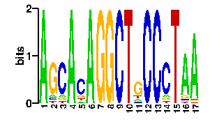
Estimated binding specificities ?
| contact |  |
A |   2   9  96  11   0   0   7  96   0   0   0   0  12   0   8   5   0
C |   2   7   0   5   0   0  70   0   0  96  96   0  12   0   5  76   0
G |   2  10   0  75  96  96  12   0  96   0   0   0  60   0  78   5   0
T |  90  70   0   5   0   0   7   0   0   0   0  96  12  96   5  10  96
scan! |
home
updated Sun Jan 4 23:48:50 2026
