
| structure name | CRYSTAL STRUCTURE OF A TWO-DOMAIN IDER-DNA COMPLEX CRYSTAL FORM I (Iron-dependent repressor ideR) |
| reference | Wisedchaisri et al. Biochemistry 46 436 2007 |
| source | Mycobacterium tuberculosis |
| experiment | X-ray (resolution=2.40, R-factor=0.201) |
| structural superfamily | Iron-dependent repressor protein, dimerization domain;"Winged helix" DNA-binding domain; |
| sequence family | Iron dependent repressor, metal bindin; Iron dependent repressor, Nterminal D; MarR family; |
| multimeric complexes | 1u8r_ABCD 1u8r_GHIJ 2isz_ABCD 2it0_ABCD |
| redundant complexes | 1u8r_D
 2it0_D 2it0_D
|
| links to other resources | NAKB  PDIdb DNAproDB PDIdb DNAproDB |
| protein sequence | |
| interface signature | SPTSQRR |
Estimated binding specificities ?
| contact | 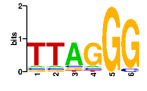 |
A |   6   9  75   6   0   0
C |   6   6   6   6   0   6
G |   9   6   6  78  96  90
T |  75  75   9   6   0   0
scan! |
Dendrogram of similar interfaces ?
matrix format--QT--PTAT--TT------------YT---- +8pw0_A +----------1 ------RGSTRTSTHG--------RA------ L ! +3zpl_B +-----3 ----------------YG-------------- ! ! +--3eyi_B +------4 +-------2 --------------RCYG----KT-------- ! ! +--2acj_B --5 ! TT------KATTQTTGLTRTKA------FTYT L ! +----------------2p7c_B ! ----DCLT--AGTTRG--RG------------ L +-----------------------5h3r_A |
home
updated Mon Jun 29 15:17:25 2026